Proximal Median Nerve Compression
Proximal Median Nerve Compression
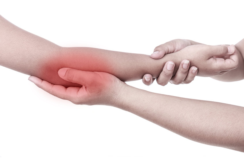
PROXIMAL MEDIAN NERVE COMPRESSION
Repetitive tasks involving: throwing, gripping, pronation (turning your hand to a palm down position), and sudden extension at the elbow can produce injury resulting in compression of the median nerve at the elbow and forearm level. Weight lifting may result in building muscle mass and can lead to nerve compression. Trauma to the upper forearm may also result in scarring and can induce compressive neuropathies at and around the elbow. Your own inherent anatomy may be a variant that makes you vulnerable to this condition. Further, it is common to be predisposed to this nerve compression if you are diabetic. Diagnosing this condition is very dependent upon the clinical evaluation and is often missed by electro-diagnostic studies ( nerve conduction, EMG).
Neuromas and Nerve Pain in General
Ives GC, Kung TA, Nghiem BT, Ursu DC, Brown DL, Cederna PS, Kemp SWP: Current State of the Surgical Treatment of Terminal Neuromas. Neurosurgery 83(3): 354-364, 2018. PM29053875
Kubiak CA, Brown DL: Principles of nerve repair and reconstruction and neuroma management. Grabb and Smith’s Plastic Surgery, Chung, Wolters Kluwer, 2019. 8th, 10, 87-96.
Hart SE, Brown DL: Dermatosensory Peripheral Nerve Interfaces: Prevention of Pain Recurrence Following Sensory Neurectomy. Hand Clin 37(3): 383-389, 2021. PM3425331
Gomez-Rexrode AE, Kennedy SH, Brown DL: Unmasked Neuropathic Pain After Neurectomy: A Case Series and Review of the Literature. Plast Reconstr Surg Glob Open.11(8): e5221, 08/2023. PM37650094
Hespe GE, Brown DL: Management of Neuropathic Pain with Neurectomy Combined with Dermal Sensory Regenerative Peripheral Nerve Interface (DS-RPNI). Semin Plas Surg. In Print.
Migraine Headaches
- Hagan R, Khansa I, Janis J. Chapter, Surgical Treatments of the Supraorbital and Supratrochlear nerves: Book, Surgical Treatment of Chronic Headaches and Migraines. March 2020.
- Scherer SS, Schiraldi L, Sapino G, Cambiaso-Daniel J, Gualdi A, Peled ZM, Hagan R, Pietramaggiori G. The Greater Occipital Nerve and Obliquus Capitis Inferior Muscle: Anatomical Interactions and Implications for Occipital Pain Syndromes. PRS Journal 2019
- Brown DL: Discussion: Efficacy of Surgical Treatment of Migraine Headaches Involving the Auriculotemporal Nerve (Site V). Plastic and Reconstructive Surgery 143(2): 564-565, PM30688902
- Hagan RR, Fallucco MA, Janis JE, Supraorbital Rim Syndrome: Definition, Surgical Treatment, and Outcomes for Frontal Headache. PRS Journal 2016.
- JanisJE, Barker JC, Javadi C, Ducic I, Hagan R, Guyuron B. A review of current evidence in the surgical treatment of migraine headaches. Plast Reconstr Surg. 2014 Oct;134(4 Suppl 2):131S-41S. doi: 10.1097
- JanisJE, Hatef DA, Hagan R, Schaub T, Liu JH, Thakar H, Bolden KM, Heller JB, Kurkjian TJ. Anatomy of the supratrochlear nerve: implications for the surgical treatment of migraine headaches. Plast Reconstr Surg. 2013 Apr;131(4):743-50.
- Vorobeichik L, Fallucco MA, Hagan RR, Chronic daily headaches secondary to greater auricular and lesser occipital neuromas following endolymphatic shunt surgery; BMJ Case Reports 2012; doi: 1136/bcr-2012-007189.
- Fallucco M, Janis J, Hagan R, The anatomical morphology of the supraorbital notch: Clinical relevance to the surgical treatment of migraine headaches; PRSJournal 2012.
Trigeminal Neuralgia
- Gualdi A, Camiaso-Daniel J, Gatti J, Peled Z, Hagan R, Bertossi D, Wurzer P, Kamolz LP, Scherer S, Pietramaggiori G. Selective denervation of the corrugator supercilia muscle for the treatment of idiopathic trigeminal neuralgia purely paroxysmal distributed in the supraorbital and supratrochlear dermatomes: The journal of Headache and pain 22:9 (2021)
Neck / Face / Shoulder Pain
- Brown DL, Dellon AL: Surgical Approach to Injuries of the Cervical Plexus and Its Peripheral Nerve Branches. Plast Reconstr Surg 141(4): 1021-1025, 2018. PM29595737
Thoracic Outlet Syndrome
- Hagan R, Ricci J, Eberlin K, Novel Surgical Approach for Decompression of the Scalene Triangle in Neuorgenic Thoracic Outlet Syndrome: J Reconstr Microsurg 2018;34:315-320.
Shoulder / Upper Extremity Pain
- Brown DL, Chung KC: Quadrangular space syndrome associated with superficial radial sensory neuropathy. Ann Plast Surg 43(2): 207-210, 1999. PM10454332
- Dhawan V, Borschel GH, Brown DL: Acute exertional compartment syndrome of the J Trauma 64(6): 1635-1637, 2008. PM16983294
Mastectomy Pain (PMPS)
- Hart S, Agarwal S, Hamill JB, Brown DL: Effective Treatment of Chronic Mastectomy Pain with Intercostal Sensory Neurectomy. Plastic and Reconstructive Surgery 14:5: 876-80, 2022.
Knee Pain
- Cinotto G, Hamill JB, Cederna PS, Kemp SWP, Brown DL: Chronic Neuropathic Knee Pain Treated with Peripheral Nerve Operations Combined with Regenerative Peripheral Nerve Interfaces. Plastic and Reconstructive Surgery.
Amputation Pain / Phantom Pain / Neuroma Pain
- Woo SL, Kung TA, Brown DL, Leonard JA, Kelly BM, Cederna PS: Regenerative Peripheral Nerve Interfaces for the Treatment of Postamputation Neuroma Pain: A Pilot Plast Reconstr Surg Glob Open 4(12): e1038, 2016. PM28293490/PMC5222635
- Hooper RC, Cederna PS, Brown DL, Haase SC, Waljee JF, Egeland BM, Kelley BP, Kung TA: Regenerative Peripheral Nerve Interfaces for the Management of Symptomatic Hand and Digital Neuromas. Plast Reconstr Surg Glob Open 8(6): e2792, 2020. PM32766027/PMC7339232
Lower Extremity / Foot Pain
- Williams E, Rosson G, Hagan R, Hashemi S, Dellon AL; Soleal Sling Syndrome (Proximal Tibial Nerve Compression): Results of Surgical Decompression; Plastic and Reconstructive Surgery Journal, Volume 129(2).
- Dellon AL, Williams E, Rosson G, Hashemi S, Tollestrup T, Hagan R, Peled Z, Gurtmueller G, Ebmer J; Denervation of the Periosteal Origin of the Adductor Muscles in Conjunction with Adductor Fasciotomy in the Surgical Treatment of Refractory Groin Pull; Plastic and Reconstructive Surgery Journal, Volume 28(4).
- Lyons DA, Brown DL: Tarsal Tunnel Neurosurgery by Example, Peripheral Nerve Volume, Selden ed; Yang and Wilson vol ed Oxford University Press, Ny, NY, 2017.
- Felder JM, Ko JH, Brown DL: Lower Extremity Contemporary Neuroma Management, Eberlein Springer, 2023. In Press.
Nerve Repair
- Brown DL, Bennett TM, Dowsing BJ, Hayes A, Abate M, Morrison WA: Immediate and delayed nerve repair: improved muscle mass and function with leukemia inhibitory J Hand Surg Am 27(6): 1048- 1055, 2002. PM12457356
Laboratory Studies
- Dhawan V, Huang Y-C, Dow D, Birla RK, Brown DL: Neurotization of in vivo tissue engineered contractile, vascularized, 3-dimensional skeletal muscle Plastic and Reconstructive Surgery 16 (3S): 179-80, 2005. 17822360
- Brown DL, Bishop DK, Wood SY, Cederna PS: Short-term anti-CD40 ligand costimulatory blockade induces tolerance to peripheral nerve allografts, resulting in improved skeletal muscle Plast Recon Surg 117(7): 2250-2258, 2006. PM16772925
- Dhawan V, Lytle IF, Dow DE, Huang YC, Brown DL: Neurotization improves contractile forces of tissue- engineered skeletal muscle. Tissue Eng. 13(11): 2813-2821, 2007. PM17822360
- Mungara AK, Brown DL, Bishop DK, Wood SY, Cederna PS: Anti-CD40L monoclonal antibody treatment induces long-term, tissue-specific, immunologic hyporesponsiveness to peripheral nerve allografts. J Reconstr Microsurg 24(3): 189-195, 2008. PM18459087
Median nerve compression in the elbow area can result in two conditions: pronator syndrome and anterior interosseus nerve syndrome. It is not uncommon for both conditions to be present at the same time.
Pronator Syndrome
Pronator syndrome produces symptoms of aching of the proximal forearm, elbow and distal arm (just above elbow). This aching may be aggravated by forceful use of the extremity, especially involving pronation. Sensory loss in the fingers and palmar components of the median nerve distribution and weakness or clumsiness in the hand are often noted, especially in the thumb and index finger.
Symptoms can be confusing with those seen in carpal tunnel syndrome. It is actually more common for proximal median nerve compression and carpal tunnel syndrome to both be present. This is considered a double crush phenomenom which is when the same nerve is pinched at more than one level along its course.
In pronator syndrome, night pain is less common (but does occur) while carpal tunnel syndrome often wakes patients with complaints of aching and numbness in the hands.
The physical examination may reveal tenderness and a Tinel sign over the pronator teres muscle. Weakness, if present, may involve the index finger, thumb flexion and pinch strength. Atrophy in the thenar muscles (base of thumb in palm) may be noted in advanced cases.
Loss or decreased sensibility of the thumb and index/long fingers are consistent with both proximal median nerve compression and carpal tunnel syndrome. Loss of sensation and/or pain in the palmar area on the thumb sites is suggestive of pronator syndrome. Provocative (exertional) tests on exam can be helpful in eliciting signs of pronator syndrome.
Anterior Interosseus Nerve Syndrome (AIN Syndrome)
The anterior interosseus nerve (AIN) syndrome as an isolated compression syndrome is more rare. AIN is a branch of the median nerve that branches from the median nerve after it passes through the pronator muscle. The AIN has no cutaneous (skin) sensory component therefore numbness is not associated with this syndrome, it is purely a motor problem.
Anterior interosseus nerve syndrome includes complaints of absent or decreased flexion (bending) of the thumb and index finger. For instance, it is difficult to make an OK sign. A weakened index finger-thumb pinch is generally noted as well.
Treatment
The initial treatment for proximal median nerve compressions is conservative. However, if severity and duration of symptoms are significant at the time of initial presentation to the physician, you may require surgery at that point.
Anterior interosseus nerve syndrome again is more rare, but usually resolves with time, particularly if it is secondary to minor trauma or irritation to the nerve. However proximal median compression in the diabetic limb usually, ultimately requires surgery and is generally found coexisting with other compressions in the same extremity.
If no improvement with time and therapy are noted or if a space occupying lesion/mass is identified on the MRI, surgical release is then recommended.
The prognosis for full recovery is generally very good, however one must consider the amount of deconditioning that was present at the time of surgery. Full recovery may take as long as 6 months even after surgical decompression. If there is severe nerve damage, recovery may take longer and may be incomplete. Risks of the surgery are very rare but include: the persistent symptoms or pain, motor weakness, nerve injury, loss of function, and other generalized surgical risks such as hematoma, seroma or neuroma.